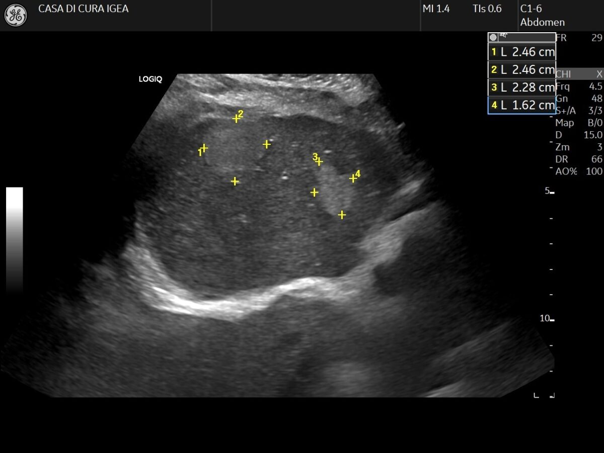
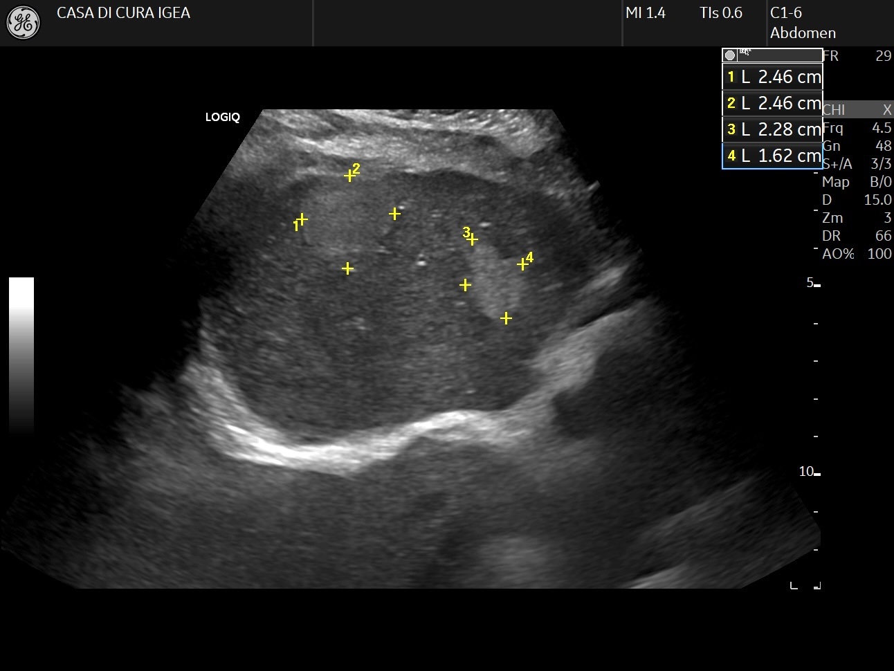
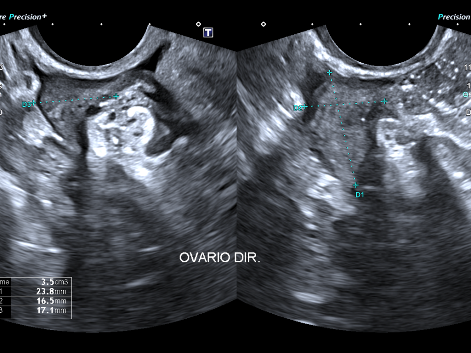
Additional images
Image 2: MRI findings of the left hepatic lesion (suspected adenoma)
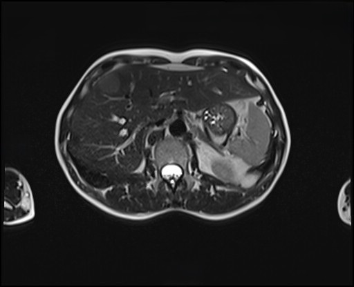
Image 2a : T2-weighted HASTE image: the lesion demonstrates subtle hyperintensity.

Image 2b : T1-weighted in and out of phase (Opposed-Phase) images: Thee out-of-phase shows a marked signal drop-off within the lesion, compatible with intracellular lipid contenent

Image 2c : Dinamic post-contrast T1-weighted VIBE images
Image 3: Dynamic post-contrast T1-weighted VIBE images
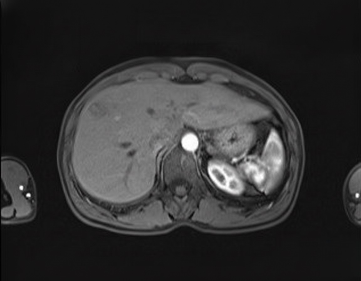
Image 3a : Arterial phase: Shows mild homogeneous enhancement
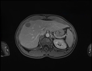
Image 3b : Portal venous phase: The lesion becomes hypointense compared to the liver parenchyma

Image 3c : Hepatobiliary phase: The lesion appears definitively hypointense, indicating a lack of functioning hepatocytes.
Findings suggestive of hepatic adenoma
Additional discussion
The hypointense appearance of HCA on the hepatobiliary phase (Image 3) is typical for most HCA subtypes, particularly the inflammatory (I-HCA) and HNF1a-inactivated (H-HCA) subtypes.
The management of HCA has evolved towards a more conservative approach for lesions <5cm in low-risk patients. The first step is always to discontinue the inciting factor, such as oral contraceptives. Subsequent management is tailored based on the lesion’s size, subtype (if available from MRI), and patient-specific risk factors, Surgical resection is typically reserved for HCAs>5cm, those that grow despite discontinuation of hormones, or specific high-risk subtypes (e.g.,Bcatenin activated). Percutaneous biopsy is not mandatory if imaging is characteristic and is often reserved for atypical cases
Conclusion
This case highlights a standard pathway for characterizing an incidental liver lesion:
- CEUS is an excellent first-line tool for dynamic vascular characterization, allowing for accurate differentiation between common benign lesions like HCA and hemangioma in a single session
- The “wash-in and wash-out” pattern in a young woman on oral contraceptives should raise strong suspicion for HCA
- MRI with hepatobiliary contrast agent is the gold standard for confirming the diagnosis, characterizing the lesion subtype, and informing risk stratification
- A multidisciplinary approach is essential for tailoring the management plan, which can range from surveillance to resection based on specific risk factors
Conflicts of Interest:
MFM : Speaker Honoraria for Bracco, General Electric
The other authors declare no conflict of interest.
References
- Dietrich CF., et al. Guidelines and Good Clinical Practice Recommendation for Contrast-Enhanced Ultrasound (CEUS) in the liver-Update 2020. Ultrashall Med 2020; 41:562-585.
- European Association for the Study of the Liver (EASL). EASL Clinical Practice Guidelines on the management of benign liver tumors. J Hepatol 2017; 65(2); 386-398
- Contrast Enhanced Ultrasound patterns of Hepatocellular Adenoma: an Italian multicenter experience. Garcovich M, Faccia M, Meloni F et al. J Ultrasound 2019. Jun;22(2):157-165
- Tse JT, Felker RE, Naini BV et al. Hepatocellular Adenomas: Molecular Basis and Multimodality Imaging Update. RadioGraphics 2023;43(3):e220134.doi:10.1148/rg.220134
- Poetter-Lang S, Ba-Ssalamah A, Hodge JC et al. Hepatocellular Adenoma Update: Diagnosis, Molecular Classification, and Clinical Course. Br J Radiol.2024 Nov 1;97(1163):1740-1754. Doi:10.1093/bjr/tqae180
Additional images
Image 2: MRI findings of the left hepatic lesion (suspected adenoma)
Image 2a : T2-weighted HASTE image: the lesion demonstrates subtle hyperintensity.
Image 2b : T1-weighted in and out of phase (Opposed-Phase) images: Thee out-of-phase shows a marked signal drop-off within the lesion, compatible with intracellular lipid contenent
Image 2c : Dinamic post-contrast T1-weighted VIBE images
Image 3: Dynamic post-contrast T1-weighted VIBE images
Image 3a : Arterial phase: Shows mild homogeneous enhancement
Image 3b : Portal venous phase: The lesion becomes hypointense compared to the liver parenchyma
Image 3c : Hepatobiliary phase: The lesion appears definitively hypointense, indicating a lack of functioning hepatocytes.
Findings suggestive of hepatic adenoma
Additional discussion
The hypointense appearance of HCA on the hepatobiliary phase (Image 3) is typical for most HCA subtypes, particularly the inflammatory (I-HCA) and HNF1a-inactivated (H-HCA) subtypes.
The management of HCA has evolved towards a more conservative approach for lesions <5cm in low-risk patients. The first step is always to discontinue the inciting factor, such as oral contraceptives. Subsequent management is tailored based on the lesion’s size, subtype (if available from MRI), and patient-specific risk factors, Surgical resection is typically reserved for HCAs>5cm, those that grow despite discontinuation of hormones, or specific high-risk subtypes (e.g.,Bcatenin activated). Percutaneous biopsy is not mandatory if imaging is characteristic and is often reserved for atypical cases
Conclusion
This case highlights a standard pathway for characterizing an incidental liver lesion:
- CEUS is an excellent first-line tool for dynamic vascular characterization, allowing for accurate differentiation between common benign lesions like HCA and hemangioma in a single session
- The “wash-in and wash-out” pattern in a young woman on oral contraceptives should raise strong suspicion for HCA
- MRI with hepatobiliary contrast agent is the gold standard for confirming the diagnosis, characterizing the lesion subtype, and informing risk stratification
- A multidisciplinary approach is essential for tailoring the management plan, which can range from surveillance to resection based on specific risk factors
Conflicts of Interest:
MFM : Speaker Honoraria for Bracco, General Electric
The other authors declare no conflict of interest.
References
- Dietrich CF., et al. Guidelines and Good Clinical Practice Recommendation for Contrast-Enhanced Ultrasound (CEUS) in the liver-Update 2020. Ultrashall Med 2020; 41:562-585.
- European Association for the Study of the Liver (EASL). EASL Clinical Practice Guidelines on the management of benign liver tumors. J Hepatol 2017; 65(2); 386-398
- Contrast Enhanced Ultrasound patterns of Hepatocellular Adenoma: an Italian multicenter experience. Garcovich M, Faccia M, Meloni F et al. J Ultrasound 2019. Jun;22(2):157-165
- Tse JT, Felker RE, Naini BV et al. Hepatocellular Adenomas: Molecular Basis and Multimodality Imaging Update. RadioGraphics 2023;43(3):e220134.doi:10.1148/rg.220134
- Poetter-Lang S, Ba-Ssalamah A, Hodge JC et al. Hepatocellular Adenoma Update: Diagnosis, Molecular Classification, and Clinical Course. Br J Radiol.2024 Nov 1;97(1163):1740-1754. Doi:10.1093/bjr/tqae180