Additional Discussion:
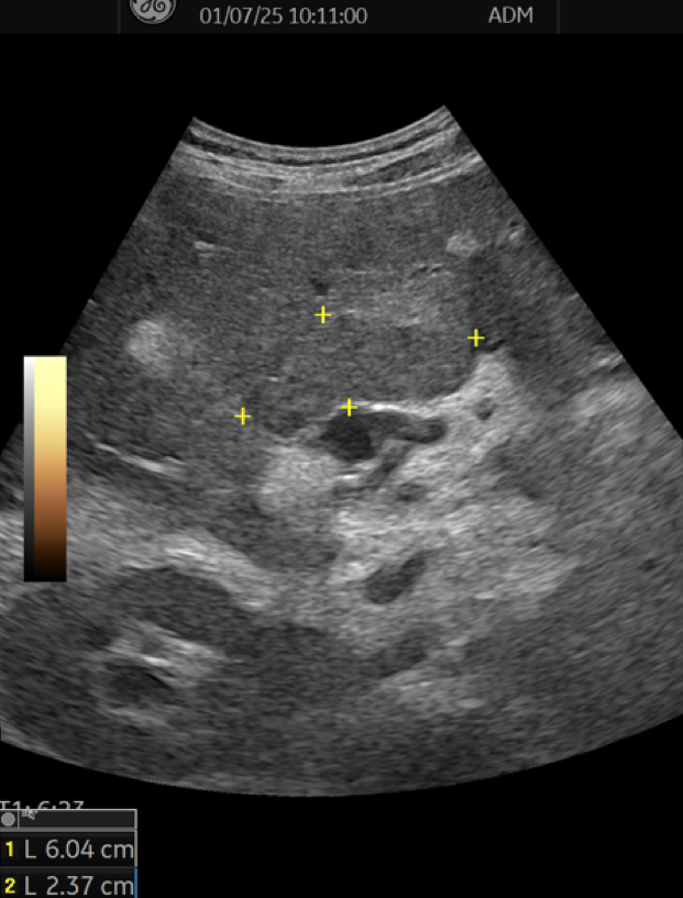
Based on the patient’s history of a prothrombotic state and liver cirrhosis, as well as the clinical, laboratory, and imaging findings, the most probable explanation for the B-mode–visible lesion is parenchymal remodeling following hypoperfusion, due to recent portal vein thrombosis.
Conclusion:
As stated by EFSUMB, contrast-enhanced ultrasound (CEUS) may serve as a first-line tool for characterizing focal liver lesions (FLLs) in patients with liver cirrhosis, aiding in the diagnosis of malignancy (CEUS LR-M) or specifically hepatocellular carcinoma (CEUS LR-5). Nevertheless, computed tomography (CT) or magnetic resonance imaging (MRI) remains necessary for precise staging, except when contraindicated (1).
Reduced hepatic vascular inflow, resulting from abnormalities of the portal vein and/or hepatic artery, can lead to a broad range of clinical outcomes. These may vary from minimal physiological impact to reversible ischemia, or to infarction, which can cause permanent liver damage or acute liver failure. On contrast-enhanced imaging, this typically appears as hypoenhancement involving part or all of the hepatic parenchyma (2). Several conditions increase the risk of vascular compromise, including hypercoagulable states, cirrhosis, pancreatitis, and a history of splenectomy, all of which predispose to the formation of bland portal vein thrombosis (3).
Given the wide variety of disorders that can affect the hepatic vasculature, it is crucial for clinicians to be familiar with their pathophysiology, imaging characteristics across modalities, and both acute and chronic presentations. Such knowledge is essential for accurate diagnosis, guiding appropriate clinical management, and assessing prognosis.
Conflicts of Interest:
“The authors declare no conflict of interest.”
References
- Dietrich CF, Nolsøe CP, et al. Guidelines and Good Clinical Practice Recommendations for Contrast-Enhanced Ultrasound (CEUS) in the Liver–Update 2020 WFUMB in Cooperation with EFSUMB, AFSUMB, AIUM, and FLAUS. Ultrasound in Medicine and Biology, Volume 46, Issue 10, 2579 – 2604.
- Ryan MF, Hamilton PA, Sarrazin J, Chu P, Benjaminov O, Lam K. The halo sign and peripancreatic fluid: useful CT signs of hypovolaemic shock complex in adults. Clin Radiol 2005;60:599-607.
- Mills A, Mellnick V. Imaging features of hepatic arterial and venous flow abnormalities. Clin Liver Dis (Hoboken). 2018 Jan 31;11(1):27-32.
Additional Discussion:
Based on the patient’s history of a prothrombotic state and liver cirrhosis, as well as the clinical, laboratory, and imaging findings, the most probable explanation for the B-mode–visible lesion is parenchymal remodeling following hypoperfusion, due to recent portal vein thrombosis.
Conclusion:
As stated by EFSUMB, contrast-enhanced ultrasound (CEUS) may serve as a first-line tool for characterizing focal liver lesions (FLLs) in patients with liver cirrhosis, aiding in the diagnosis of malignancy (CEUS LR-M) or specifically hepatocellular carcinoma (CEUS LR-5). Nevertheless, computed tomography (CT) or magnetic resonance imaging (MRI) remains necessary for precise staging, except when contraindicated (1).
Reduced hepatic vascular inflow, resulting from abnormalities of the portal vein and/or hepatic artery, can lead to a broad range of clinical outcomes. These may vary from minimal physiological impact to reversible ischemia, or to infarction, which can cause permanent liver damage or acute liver failure. On contrast-enhanced imaging, this typically appears as hypoenhancement involving part or all of the hepatic parenchyma (2). Several conditions increase the risk of vascular compromise, including hypercoagulable states, cirrhosis, pancreatitis, and a history of splenectomy, all of which predispose to the formation of bland portal vein thrombosis (3).
Given the wide variety of disorders that can affect the hepatic vasculature, it is crucial for clinicians to be familiar with their pathophysiology, imaging characteristics across modalities, and both acute and chronic presentations. Such knowledge is essential for accurate diagnosis, guiding appropriate clinical management, and assessing prognosis.
Conflicts of Interest:
“The authors declare no conflict of interest.”
References
- Dietrich CF, Nolsøe CP, et al. Guidelines and Good Clinical Practice Recommendations for Contrast-Enhanced Ultrasound (CEUS) in the Liver–Update 2020 WFUMB in Cooperation with EFSUMB, AFSUMB, AIUM, and FLAUS. Ultrasound in Medicine and Biology, Volume 46, Issue 10, 2579 – 2604.
- Ryan MF, Hamilton PA, Sarrazin J, Chu P, Benjaminov O, Lam K. The halo sign and peripancreatic fluid: useful CT signs of hypovolaemic shock complex in adults. Clin Radiol 2005;60:599-607.
- Mills A, Mellnick V. Imaging features of hepatic arterial and venous flow abnormalities. Clin Liver Dis (Hoboken). 2018 Jan 31;11(1):27-32.