An Incidental Liver Lesion: Is Biopsy Always Necessary?
Marta Columbu[1], Debora Cidoni[4], Mariangela Fortunato[3], Paolo Buscemi[2], Maria Franca Meloni[5]
[1] Department of Radiology, Local Health Authority (ASL) Cagliari, SS Hospital Trinità, Cagliari, Italy
[2] Department of Interventional Radiology Unit, ARNAS Civico, Palermo, Italy
[3] Department of Biomedical Sciences, Humanitas University, Pieve Emanuele Milano , Italy
[4] Department of Radiology,ASPRC, Locri, Italy
[5] Department of Radiology , Casa di Cura Igea, Milano , Italy
Corresponding author: meloni.mariafranca@gmail.com
Clinical history
A 72-year-old physically active man in excellent general condition, with no significant comorbidities and negative serology for hepatotropic viral infections (HBV and HCV), was a former blood donor. His medical history included hepatic steatosis and indirect hyperbilirubinemia related to known Gilbert’s syndrome. He underwent annual abdominal ultrasound follow-up for a 4 cm right renal cyst classified as Bosniak II. During the most recent ultrasound examination, a 4.9 × 4.7 cm focal liver lesion was incidentally detected in segment VI, associated with portal vein thrombosis. The patient was asymptomatic.
Ultrasound Images
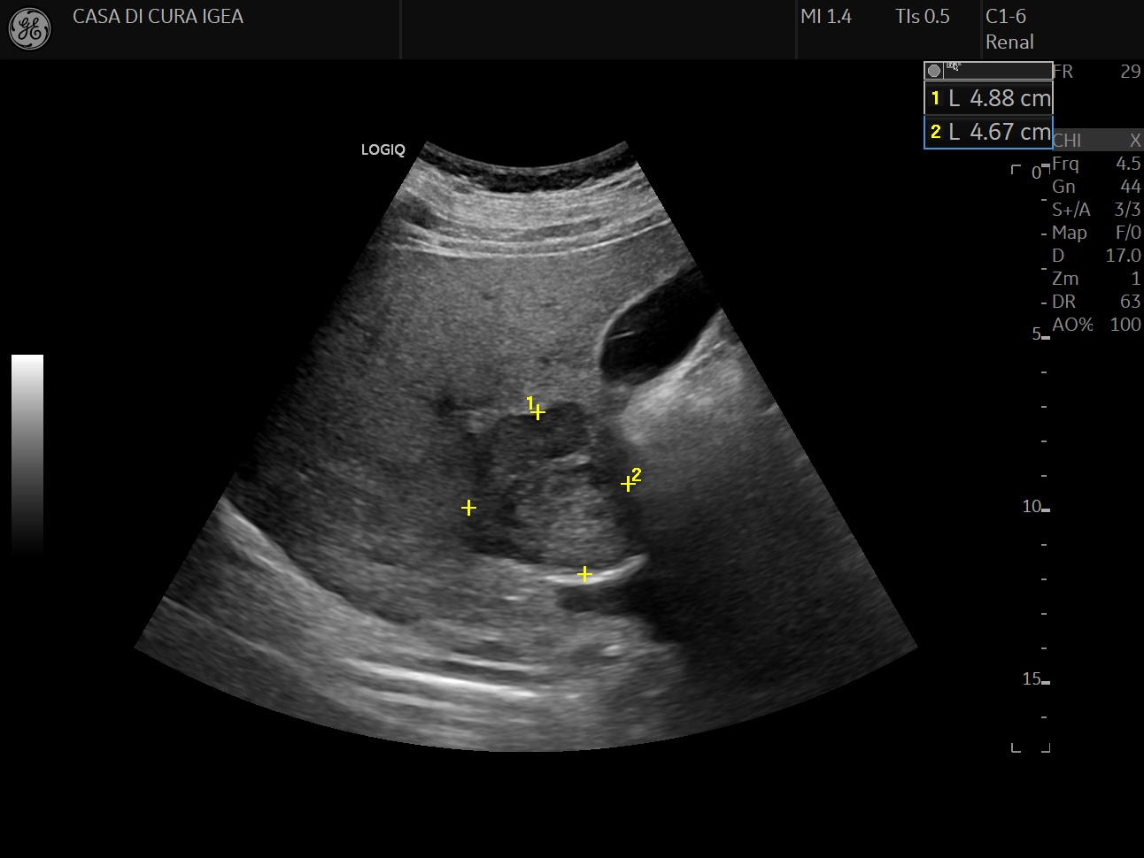
Image 1. B-mode ultrasound of the liver shows a large, well-defined focal lesion in segment VI, measuring approximately 4.9 × 4.7 cm in this scan plane. The lesion is hypoechoic, with heterogeneous internal echotexture, irregular margins, and a visible peripheral halo. It is located close to the liver capsule and adjacent portal vein branches. No significant posterior acoustic enhancement is observed.
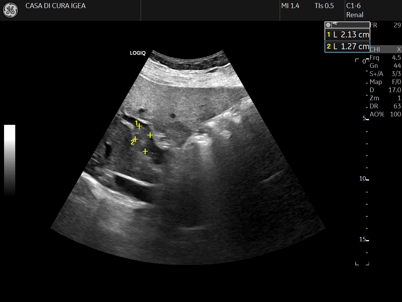
Image 2. B-mode ultrasound of the liver shows a tubular structure containing echogenic material within its lumen, consistent with a thrombus partially occluding the vessel. The thrombus measures approximately 2.1 × 1.3 cm in this scan plane. The surrounding liver parenchyma shows mild steatosis.
Following incidental detection of the focal liver lesion on B-mode ultrasound, contrast-enhanced ultrasound (CEUS) was performed in accordance with EFSUMB guidelines for the characterisation of focal liver lesions.
Video 1. CEUS (contrast-enhanced ultrasound) of the liver lesion.
The video demonstrates the complete contrast-enhancement pattern. During the arterial phase (19–25 s), the lesion shows rapid and intense arterial-phase hyperenhancement, appearing markedly hyperechoic relative to the surrounding liver parenchyma. In the portal venous phase (56 s), the lesion begins to show wash-out, becoming progressively isoechoic relative to the surrounding liver.
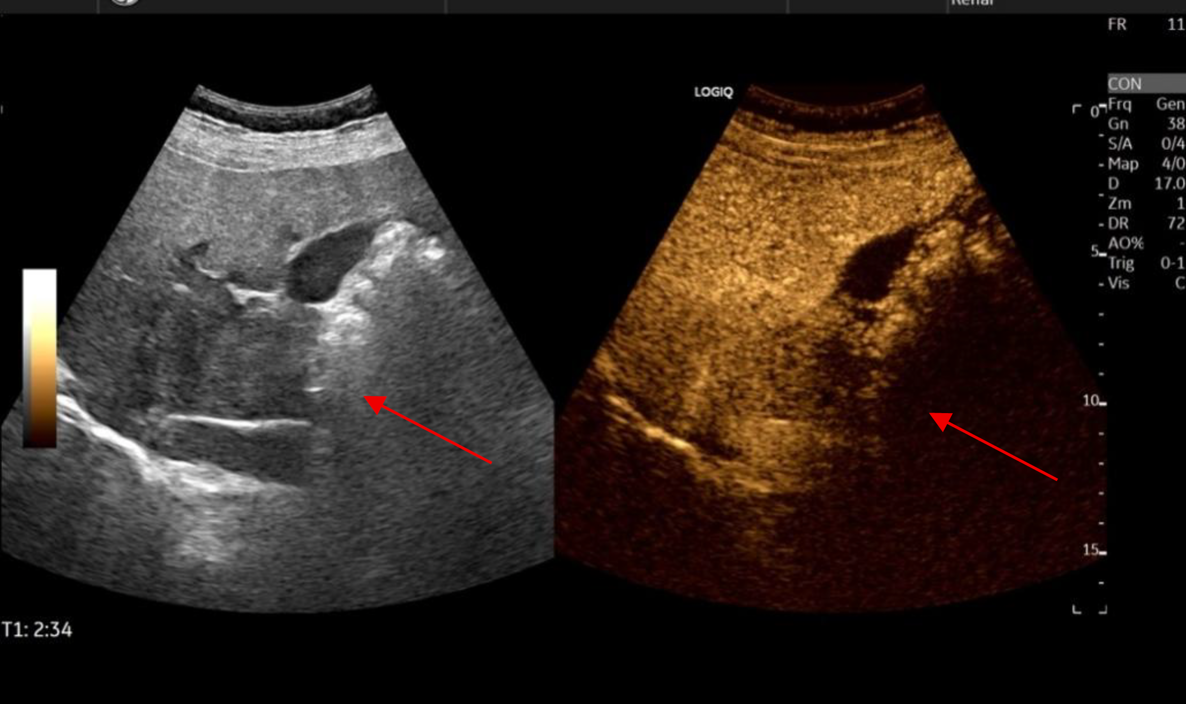
Image 3. CEUS of the liver lesion, late phase (2:34 min).
The lesion appears hypoechoic relative to thesurrounding liver parenchyma, consistent with progressive wash-out.
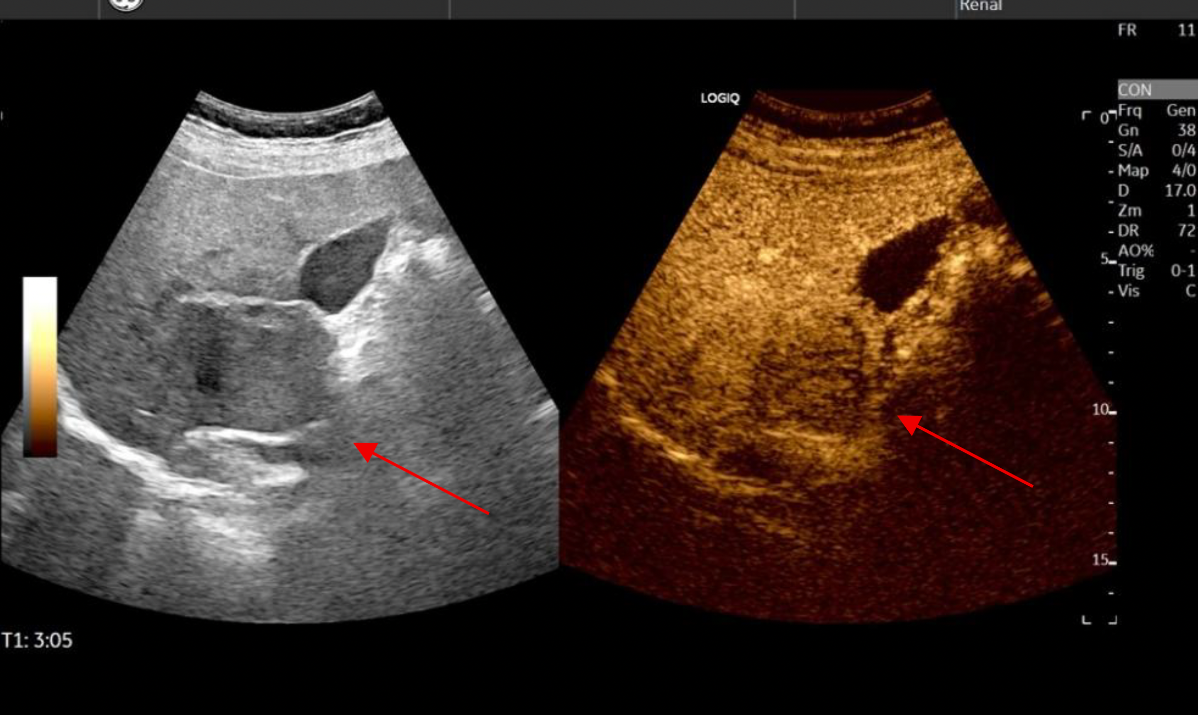
Image 4. CEUS of the liver lesion, late phase (3:05 min).
The lesion appears markedly hypoechoic (“black spot”) relative to the enhancing liver parenchyma, consistent with complete late wash-out. CEUS was also used to assess the portal vein, where thrombus was identified. The enhancement pattern of the thrombus was evaluated and compared with that of the liver lesion.
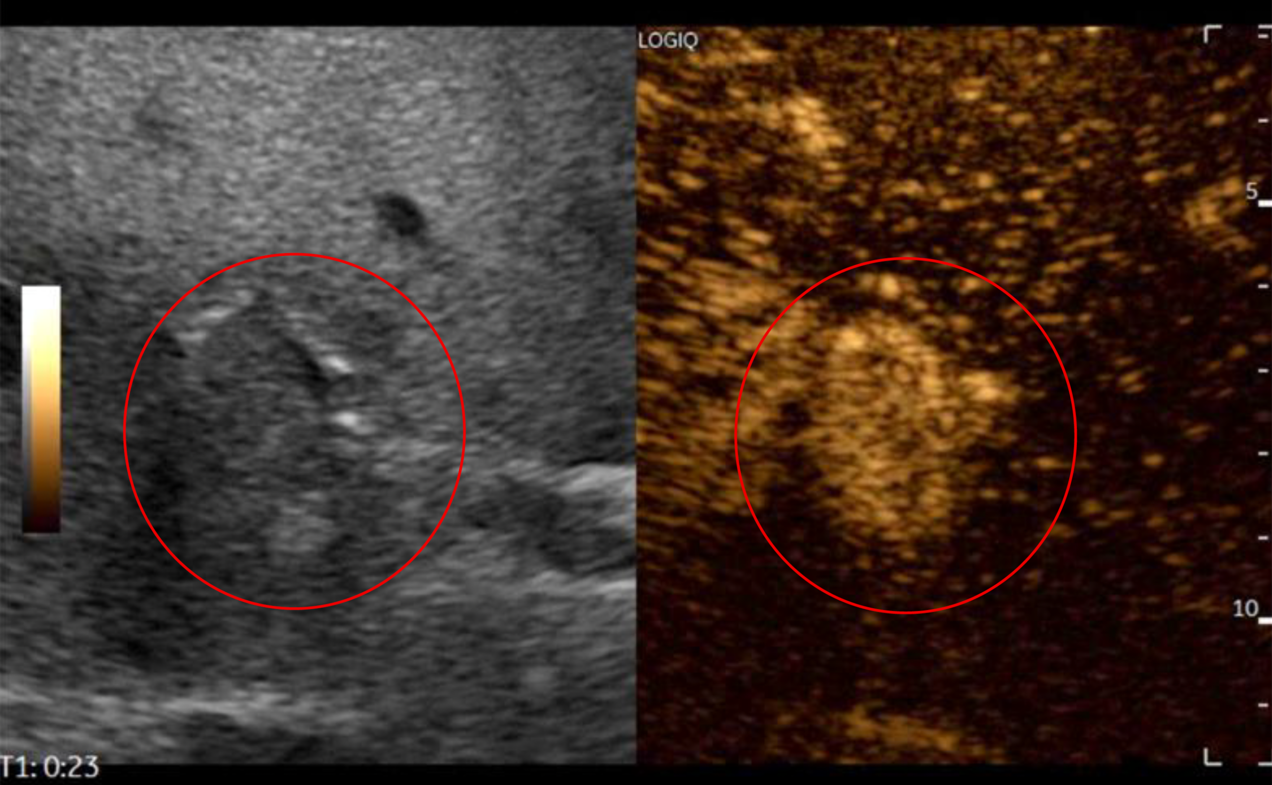
Image 5. CEUS of the portal vein thrombus, arterial phase (23 s). The thrombus shows contrast enhancement, appearing hyperechoic within the vessel lumen. This finding indicates arterial vascularization of the thrombus.
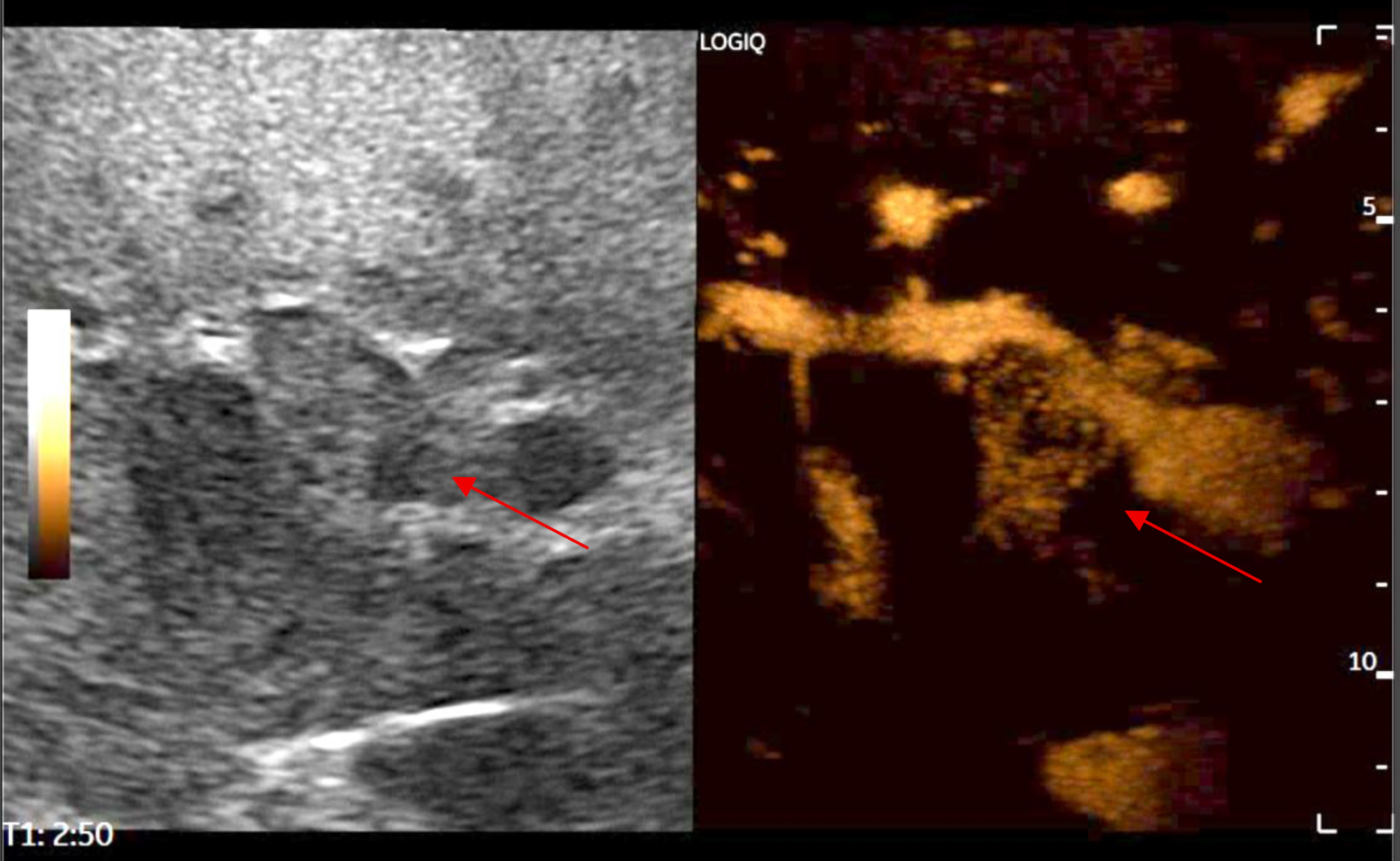
Image 6. CEUS of the portal vein thrombus, late phase after flash capture (2:50 min). Following flash capture, microbubbles within the scan plane are destroyed, producing a transient dark appearance of the surrounding liver parenchyma. The thrombus appears completely devoid of contrast enhancement, consistent with complete wash-out. The contrast-enhancement pattern of the thrombus is superimposable on that of the liver lesion observed in the arterial and late phases.