Unusual Solid Pancreatic Lesion in an Asymptomatic Patient
Maria Franca Meloni(1), Marta Columbu(2), Paolo Buscemi(3), and Debora Cidoni
(4)
(1) Casa di Cura Igea, Department of Radiology, Milan, Italy
(2) Department of Radiology, Local Health Authority (ASL) Cagliari, SS Hospital. Trinità, Cagliari, Italy
(3) Interventional Radiology Unit, ARNAS Civico, Palermo, Italy
(4) Department of Radiology, Provincial Health Authority of Reggio Calabria, Locri, Italy
* Correspondence: meloni.mariafranca@gmail.com
Clinical history
A 58-year-old asymptomatic male with no significant medical history underwent an abdominal MRI for follow-up of a known simple pancreatic cyst. Incidentally, the MRI revealed a new, well-defined, round, solid lesion measuring 1.1 cm in the tail of the pancreas, separate from the known cyst.
No dilatation of the pancreatic duct or perilesional abnormalities were observed.
Laboratory tests, including serum amylase, lipase, and chromogranin A, were within normal limits.
MRI Images
Image 1: MRI findings pre-contrast
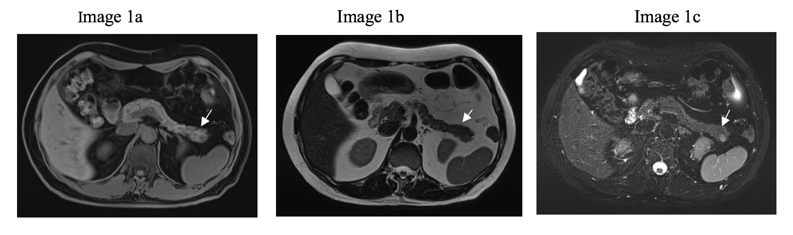
1a: T1-vibe: At the dorsal aspect of the pancreatic tail, an oval-shaped lesion is observed, appearing hypointense and mildly heterogeneous compared with the surrounding pancreatic parenchyma on T1-weighted images.
1b: T2-TSE: On T2-weighted sequences, the oval-shaped lesion appears isointense relative to the adjacent pancreatic parenchyma.
1c: T2-FS: On fat-suppressed T2-weighted sequences, the oval-shaped lesion appears hyperintense.

1d-e: DWI: On diffusion-weighted imaging (DWI), the oval lesion demonstrates mild signal restriction, similar to the physiological behavior of splenic parenchyma, with a corresponding high signal on the ADC map, indicating a “shine-through “effect (arrows)
MRI Images
Image 2: Dynamic post-contrast (gadobutrol) T1-weighted VIBE images:

2a: Arterial phase; 2b: Portal venous phase; 2c: Late phase.
Following intravenous administration of the contrast agent, the oval-shaped lesion demonstrates mild and heterogeneous enhancement throughout all dynamic phases (arterial, portal venous, and delayed), without evidence of washout, showing a progressive tendency toward isointensity relative to the surrounding pancreatic parenchyma.
Video 1 (CEUS- Arterial to Late phase) : Dynamic cine loop from 15 seconds to 10 minutes post-contrast injection. The pancreatic lesion (arrow) demonstrates intense, homogeneous enhancement synchronous with the splenic parenchyma in the early arterial phase persisting identically without wash-out throughout the observation period (up to 10 minutes after injection)