Discussion
Ultrasound reveals an incisional hernia due to a 1 cm defect in the abdominal wall located at one of the port sites from the recent laparotomy. The hernia sac measures about 3x1 cm in diameter.
Hematomas and abscesses are also potential post-surgical complications; however, the visualized defect (red arrows) indicates a hernia (Figure 3). Also, the physical examination with no palpable pain makes hematoma and abscess less likely. If in doubt, Doppler imaging can potentially be helpful in finding the correct diagnosis. The contents of a hernia usually have their own blood supply, which can often be visualized by color Doppler. Furthermore, hematoma, abscesses, and seroma are usually not moveable with the transducer and an intraabdominal connection is usually not seen.
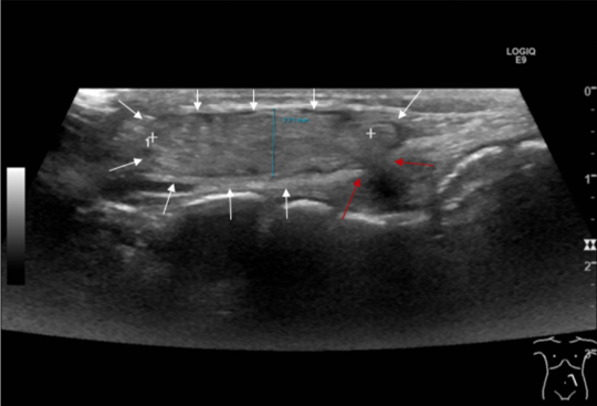
Figure 2: Same image as in figure 1. The white arrows point to the hernial sac. The red arrows indicate the defect in the abdominal wall.
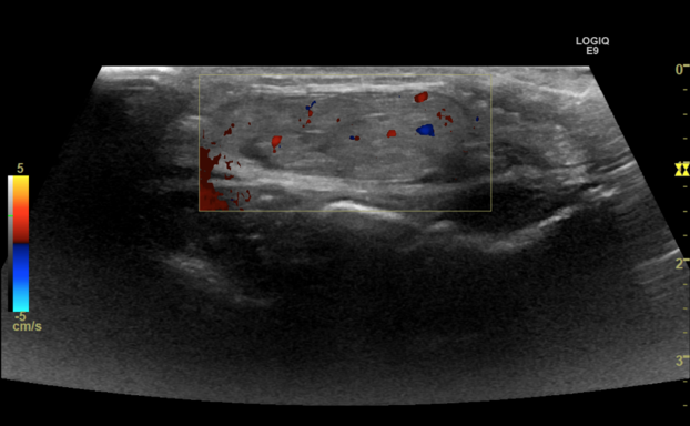
Figure 3: Same image as in figure 1 with color Doppler.
The referring surgeon requested to know the content of the hernia sac. What would you answer?
Additional discussion
The diagnosis is made by looking at the morphology of the contents in the hernia sac. Bowel will contain a lumen, while omental fat will not. Strangulated bowel or strangulated omental fat in a hernia should be suspected in the case of absent color Doppler signal. If strangulated bowel in a hernia is suspected, immediate surgery is essential to prevent bowel compromise or loss.
An incisional hernia is the bulging of abdominal contents through a surgical or traumatic defect in the abdominal wall's muscle and facia layers. (1). Not much is known about how often incisional hernias occur in children. One Japanese retrospective study found an incidence of 0.68% (14/2049) among children who had undergone abdominal laparotomy or laparoscopy, indicating it as a rare complication. (2).
The diagnosis is often established clinically; however, imaging is needed when clinical findings are inconclusive, complications are suspected, or surgical repair is considered. Ultrasound offers a cost-effective, real-time assessment with no ionizing radiation, rendering it particularly appropriate for pediatric patients. It enables evaluation of the hernia sac contents and measurement of the abdominal defect. (3). Incisional hernias in children often need surgical repair to strengthen the weakened area and prevent further complications like incarceration and strangulation. (3).
Conclusion
Ultrasound is a fast and safe tool to evaluate postsurgical superficial abdominal complications in children.
Conflicts of Interest:
Declare conflicts of interest or state “The authors declare no conflict of interest.”
References
- Korenkov M, Paul A, Sauerland S, et al. Classification and surgical treatment of incisional hernia. Results of an experts' meeting. Arch Surg. 2001;386:65–73.
- Tanaka, Misawa, et al. Risk Factors for Incisional Hernia in Children. World J Surg. 2018 Jul;42(7):2265-2268
- Parikh KR, Al-Hawary M, Millet JD, Burney R, Finks J, Maturen K. Incisional Hernia Repair: What the Radiologist Needs to Know. AJR Am J Roentgenol. 2017 Dec;209(6):1239-1246.
Additional discussion
The diagnosis is made by looking at the morphology of the contents in the hernia sac. Bowel will contain a lumen, while omental fat will not. Strangulated bowel or strangulated omental fat in a hernia should be suspected in the case of absent color Doppler signal. If strangulated bowel in a hernia is suspected, immediate surgery is essential to prevent bowel compromise or loss.
An incisional hernia is the bulging of abdominal contents through a surgical or traumatic defect in the abdominal wall's muscle and facia layers. (1). Not much is known about how often incisional hernias occur in children. One Japanese retrospective study found an incidence of 0.68% (14/2049) among children who had undergone abdominal laparotomy or laparoscopy, indicating it as a rare complication. (2).
The diagnosis is often established clinically; however, imaging is needed when clinical findings are inconclusive, complications are suspected, or surgical repair is considered. Ultrasound offers a cost-effective, real-time assessment with no ionizing radiation, rendering it particularly appropriate for pediatric patients. It enables evaluation of the hernia sac contents and measurement of the abdominal defect. (3). Incisional hernias in children often need surgical repair to strengthen the weakened area and prevent further complications like incarceration and strangulation. (3).
Conclusion
Ultrasound is a fast and safe tool to evaluate postsurgical superficial abdominal complications in children.
Conflicts of Interest:
Declare conflicts of interest or state “The authors declare no conflict of interest.”
References
- Korenkov M, Paul A, Sauerland S, et al. Classification and surgical treatment of incisional hernia. Results of an experts' meeting. Arch Surg. 2001;386:65–73.
- Tanaka, Misawa, et al. Risk Factors for Incisional Hernia in Children. World J Surg. 2018 Jul;42(7):2265-2268
- Parikh KR, Al-Hawary M, Millet JD, Burney R, Finks J, Maturen K. Incisional Hernia Repair: What the Radiologist Needs to Know. AJR Am J Roentgenol. 2017 Dec;209(6):1239-1246.