DISCUSSION:
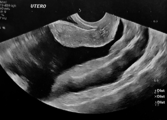
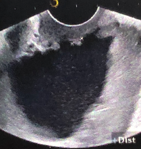
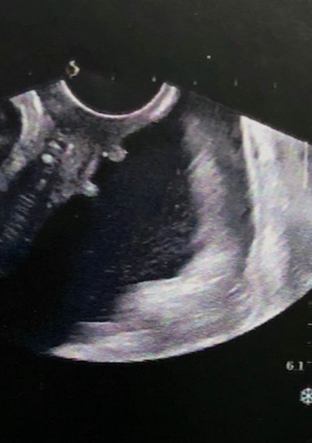
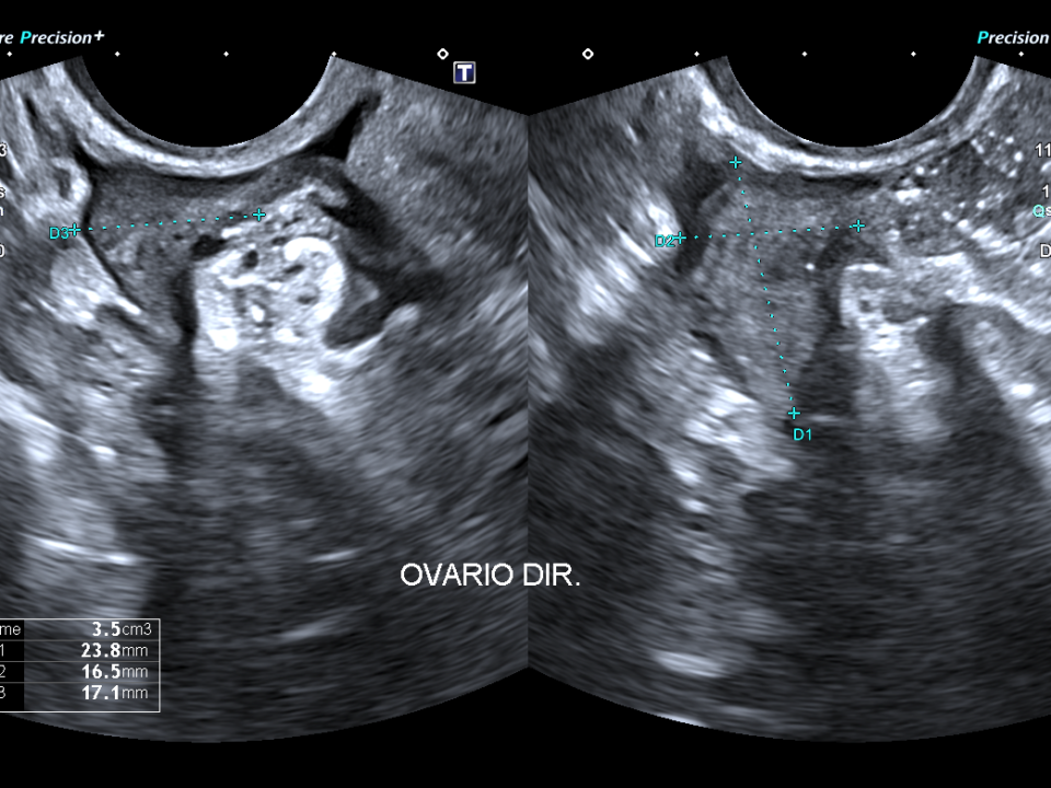
The ascitic fluid study confirmed ovarian neoplasia, which was not visible with supraapubic or transvaginal ultrasound, nor with MRI.
Since schistosomiasis mansoni causes periportal fibrosis, it is a pre-sinusoidal disease, the ascites produced is minor. The patient does indeed present periportal fibrosis on abdominal ultrasound, but the amount of ascites suggests that schistosomiasis mansoni is not the etiology, given the volume, and other pathologies should be considered.
Schistosomiasis mansoni usually presents with increase in size of the left hepatic lobe, right lobe atrophy, periportal fibrosis, small ascites and very pronounced splenomegaly, unlike chronic parenchymal liver diseases of non-schistosomal etiology, which does not present with localized increase in the left lobe, right lobe atrophy, does not present periportal fibrosis, can present with significant amount of ascites and splenomegaly is less pronounced.
CONCLUSION:
During our training, we are taught to look for a single disease that explains all of a patient's findings. However, although uncommon, the coexistence of two separate conditions is possible. When imaging findings do not align with the patient's known diagnosis, we must consider this possibility. While rare, it does happen.
BIBLIOGRAPHY:
1. Ultrassonografia geral / editor da coleção Ronaldo Hueb Baroni ; editores do volume Antonio Carlos Matteoni de Athayde, Leticia Martins Azeredo. São Paulo : Editora dos Editores, 2024. -- (Coleção CBR : Radiologia e Diagnóstico por imagem ; 14), ISBN 978-65-6103-040-3
2. Pinto-Silva RA, Queiroz LC, Azeredo LM, Silva LC, Lambertucci JR. Ultrasound in schistosomiasis mansoni. Mem Inst Oswaldo Cruz. 2010 Jul;105(4):479-84. doi: 10.1590/s0074-02762010000400021. PMID: 20721494
3. Cerri GG, Alves VA, Magalhães A. Hepatosplenic schistosomiasis mansoni: ultrasound manifestations. Radiology. 1984 Dec;153(3):777-80. doi: 10.1148/radiology.153.3.6387793. PMID: 6387793
4. Ultra-sonografia Abdominal, Segunda Edição, Maria Cristina Chammas, Giovani Guido Cerri, 2009, ISBN 978-85-372-0186-2
DISCUSSION:
The ascitic fluid study confirmed ovarian neoplasia, which was not visible with supraapubic or transvaginal ultrasound, nor with MRI.
Since schistosomiasis mansoni causes periportal fibrosis, it is a pre-sinusoidal disease, the ascites produced is minor. The patient does indeed present periportal fibrosis on abdominal ultrasound, but the amount of ascites suggests that schistosomiasis mansoni is not the etiology, given the volume, and other pathologies should be considered.
Schistosomiasis mansoni usually presents with increase in size of the left hepatic lobe, right lobe atrophy, periportal fibrosis, small ascites and very pronounced splenomegaly, unlike chronic parenchymal liver diseases of non-schistosomal etiology, which does not present with localized increase in the left lobe, right lobe atrophy, does not present periportal fibrosis, can present with significant amount of ascites and splenomegaly is less pronounced.
CONCLUSION:
During our training, we are taught to look for a single disease that explains all of a patient's findings. However, although uncommon, the coexistence of two separate conditions is possible. When imaging findings do not align with the patient's known diagnosis, we must consider this possibility. While rare, it does happen.
BIBLIOGRAPHY:
1. Ultrassonografia geral / editor da coleção Ronaldo Hueb Baroni ; editores do volume Antonio Carlos Matteoni de Athayde, Leticia Martins Azeredo. São Paulo : Editora dos Editores, 2024. -- (Coleção CBR : Radiologia e Diagnóstico por imagem ; 14), ISBN 978-65-6103-040-3
2. Pinto-Silva RA, Queiroz LC, Azeredo LM, Silva LC, Lambertucci JR. Ultrasound in schistosomiasis mansoni. Mem Inst Oswaldo Cruz. 2010 Jul;105(4):479-84. doi: 10.1590/s0074-02762010000400021. PMID: 20721494
3. Cerri GG, Alves VA, Magalhães A. Hepatosplenic schistosomiasis mansoni: ultrasound manifestations. Radiology. 1984 Dec;153(3):777-80. doi: 10.1148/radiology.153.3.6387793. PMID: 6387793
4. Ultra-sonografia Abdominal, Segunda Edição, Maria Cristina Chammas, Giovani Guido Cerri, 2009, ISBN 978-85-372-0186-2