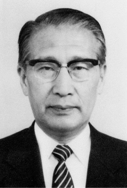
In Memoriam: Morimichi Fukuda (1929 – 2021)
Mourning the death of Dr. Morimichi Fukuda (Japan)
Dr. Morimichi Fukuda passed away on April 1, 2021. We would like to express our deepest condolences and express our sincere gratitude.
Here, I would like to show you a part of Professor Fukuda’s career, look back on his achievements, remember the deceased, and pray for his soul. Professor Fukuda was born in Sapporo in June 1929. After graduating from Hokkaido University School of Medicine in 1953 and completing a medical practice (internship) at the Sapporo Medical University Hospital in 1954, he became a research assistant in the Department of Internal Medicine, Sapporo Medical University and the Clinical Division of the Cancer Institute, and then became an assistant professor. From 1958 to 1960, he conducted research on “cancer” immunity in UCLA, California, USA. He conducted biochemical research on Cancer Cachexia, and also served as a clinician at the Department of Internal Medicine, Cancer Institute, Sapporo Medical University.
Development of ultrasonic laparoscope and ultrasonic endoscope: Spatial resolution by high frequency probe that breaks the limit in the process of comparative research between diagnosis of hepatocellular carcinoma from the body surface and pathological diagnosis He felt the need to observe and record the lesion from a short distance in order to obtain a good ultrasonic image, and worked on the development of an ultrasonic laparoscope. In addition, he aimed to observe upper gastrointestinal mucosal lesions, submucosal tumors, and lesions of organs in contact with the upper gastrointestinal tract from a short distance with a high-frequency probe using high-resolution ultrasonic images. He has developed an intraluminal ultrasound diagnostic device equipped with a child and have achieved the practical application of an ultrasonic endoscope equipped with an optical endoscope. This transmucosal endoscopic echography method is used not only for the abdominal region such as submucosal invasion of early gastric
cancer, submucosal tumor, adjacent hepatobiliary and pancreatic lesions, abdominal cavity, superior intestinal tract, and inferior intestinal artery lesions, but also for circulation. It is widely used for transesophageal ultrasonography such as left atrioventricular lesions in the organ region and transesophageal ultrasonography for mediastinal lesions in the respiratory region, but it can be said to be one of the studies that opened the beginning.
Multicenter interpretation experiment and objective evaluation method of interpretation ability: Real-time automatic with eight large-diameter long-focus probes developed by Dr. Kosoff for manually scanned ultrasound images that often depend on the diagnostic technique of the examiner. We examined the clinical usefulness of the scanning ultrasonic diagnostic device “Octoson” . Although there is a limit to the size of the scanning cross section, real-time automatic scanning is realized by an electronic scanning ultrasonic diagnostic device, and the subsequent application to the device and its multi-organs is based on X-ray images, CT images, ultrasonic images, etc. The project of Ministry of Health, Labor and Welfare, “Research on multiple image diagnosis of cancer” was taken over by the subsidy research group (Fukuda group), and in addition to the development of diagnostic equipment, multicenter research based on image interpretation experiments was conducted, and differences between image facilities and image interpretation methods. He has paved the way for standardization of diagnostic imaging by disseminating objective evaluations of individual differences in image interpretation ability.
He was Chairman of the International Exchange Committee of the Japan Society of Ultrasonics in Medicine in 1988-1992, Vice-Chairman in 1992-1994, and a member in 1996-1998. During this time, he was the President of WFUMB in 1991-1994. WFUMB94 Sapporo was held as the chairman of the congress meeting in July 2014.
Development of ultrasound diagnosis and ultrasound endoscopy of the abdominal region, establishment of an objective evaluation method for cancer image diagnosis using CT images and ultrasound images by an interpretation doctor at another facility. After retiring from Sapporo Medical University, he was active as an honorary professor and trained younger generations. We believe that these achievements will remain in the future education, clinical research, and social activities of many people who have been influenced by his activities.
Written by Professor Emeritus, Sapporo Medical University, Hiroshi Natori, Translated by Iwaki Akiyama
More can be found on the UMB Journal at https://www.umbjournal.org/article/S0301-5629(21)00302-1/fulltext